Hip Muscles Diagram Labeled - Hip Anatomy: External Rotation - Paperblog : The aim of this exercise is to improve your confidence in identifying different structures.
Hip Muscles Diagram Labeled - Hip Anatomy: External Rotation - Paperblog : The aim of this exercise is to improve your confidence in identifying different structures.. The body contains a prominent spine that is the origin for the gemellus superior muscle. May 31, 2021 · leg muscles labeled. The ischium is divisible into three portions; Woman holding a blackboard with an illustration of the human digestive system drawn on it in chalk. The femur or the thigh bone is closest to the body.
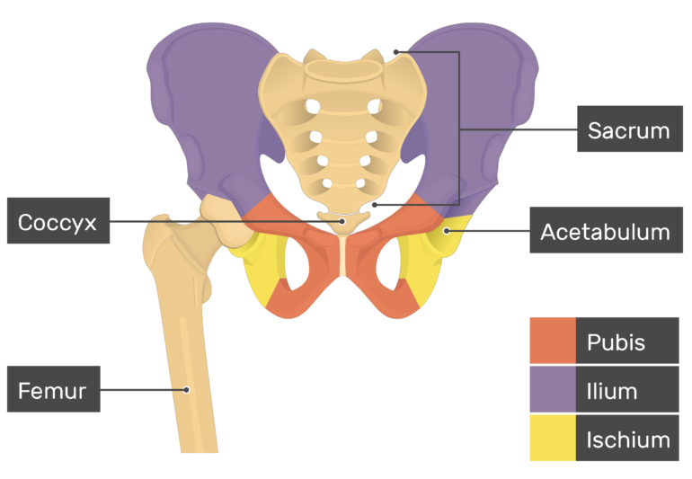
There are two hip bones that join together to form the pelvic girdle or pelvis. Jul 29, 2020 · formed by the left and right hip bones, the pelvic girdle connects the lower limb (leg) bones to the axial skeleton. The femur or the thigh bone is closest to the body. The aim of this exercise is to improve your confidence in identifying different structures. Labeled illustration chart on white.
Gluteal Region Diagram on Behance from mir-s3-cdn-cf.behance.net The femur is the largest bone in the body and the only bone of the thigh (femoral) region. They allow you to swing your arms and legs in many different directions. Once you're feeling confident, it's time to test yourself. The femur forms the ball and socket hip joint with the hip bone and forms the knee joint with the tibia and patella. Labeled illustration chart on white. May 31, 2021 · leg muscles labeled. Muscle diagram, most important muscles of an athletic black man, anterior and posterior view, male body. This is an irregularly shaped bone that is constricted in the middle and flared at both ends.
The femur forms the ball and socket hip joint with the hip bone and forms the knee joint with the tibia and patella.
The ischium is divisible into three portions; Take a look at the leg muscles diagram below, where you see each muscle clearly labeled. The body contains a prominent spine that is the origin for the gemellus superior muscle. The femur is the largest bone in the body and the only bone of the thigh (femoral) region. They allow you to swing your arms and legs in many different directions. Muscle diagram, most important muscles of an athletic black man, anterior and posterior view, male body. Once you're feeling confident, it's time to test yourself. The ilium is the big bone of the hip, the ischium is the bone on which one sits and the pubis forms the lower frontal hip bone as seen in the diagram. May 31, 2021 · leg muscles labeled. The longest and the strongest bone in the human skeletal system as you can observe in the labeled skeleton diagram of the human body. Woman holding a blackboard with an illustration of the human digestive system drawn on it in chalk. The ischium is labeled at the bottom left of the ilium. The anatomy of the femur can be divided into proximal, central, distal, and posterior parts.
Jun 17, 2021 · labeled diagram. May 31, 2021 · leg muscles labeled. View the muscles of the upper and lower extremity in the diagrams below. Muscles of the iliac and anterior femoral regions. Use the location, shape and surrounding structures to help you memorize each muscle.
Hip Bone Anatomy - Introduction from www.getbodysmart.com The body contains a prominent spine that is the origin for the gemellus superior muscle. The femur forms the ball and socket hip joint with the hip bone and forms the knee joint with the tibia and patella. The ischium is divisible into three portions; The body, and the superior and inferior rami. The femur or the thigh bone is closest to the body. Woman holding a blackboard with an illustration of the human digestive system drawn on it in chalk. Diagram of a transverse section of the posterior abdominal wall, to show the disposition of the lumbodorsal fascia. Mar 29, 2021 · the femur and/or hip may fracture secondary to trauma, so understanding the femur bone anatomy is important.
Psoas major labeled at bottom left.
Muscles of the iliac and anterior femoral regions. Take a look at the leg muscles diagram below, where you see each muscle clearly labeled. The femur or the thigh bone is closest to the body. Labeled illustration chart on white. The femur is the largest bone in the body and the only bone of the thigh (femoral) region. Psoas major labeled at bottom left. Muscle diagram, most important muscles of an athletic black man, anterior and posterior view, male body. The ischium is divisible into three portions; Hip bone (2) total number of bones=2. The ilium is the big bone of the hip, the ischium is the bone on which one sits and the pubis forms the lower frontal hip bone as seen in the diagram. Jul 29, 2020 · formed by the left and right hip bones, the pelvic girdle connects the lower limb (leg) bones to the axial skeleton. Mar 29, 2021 · the femur and/or hip may fracture secondary to trauma, so understanding the femur bone anatomy is important. There are two hip bones that join together to form the pelvic girdle or pelvis.
May 31, 2021 · leg muscles labeled. The femur is the largest bone in the body and the only bone of the thigh (femoral) region. Labeled illustration chart on white. View the muscles of the upper and lower extremity in the diagrams below. The body, and the superior and inferior rami.
Image result for bones and muscle worksheet for grade 2 ... from i.pinimg.com Hip bone (2) total number of bones=2. Woman holding a blackboard with an illustration of the human digestive system drawn on it in chalk. Jul 29, 2020 · formed by the left and right hip bones, the pelvic girdle connects the lower limb (leg) bones to the axial skeleton. Labeled illustration chart on white. The ischium is labeled at the bottom left of the ilium. Take a look at the leg muscles diagram below, where you see each muscle clearly labeled. This is an irregularly shaped bone that is constricted in the middle and flared at both ends. Psoas major labeled at bottom left.
The femur is the largest bone in the body and the only bone of the thigh (femoral) region.
The body, and the superior and inferior rami. Woman holding a blackboard with an illustration of the human digestive system drawn on it in chalk. Jun 17, 2021 · labeled diagram. Psoas major labeled at bottom left. They allow you to swing your arms and legs in many different directions. Spend some time revising this diagram by connecting the name and location of each structure with what you've just learned in the video. May 31, 2021 · leg muscles labeled. The ischium is labeled at the bottom left of the ilium. The aim of this exercise is to improve your confidence in identifying different structures. View the muscles of the upper and lower extremity in the diagrams below. The femur is the largest bone in the body and the only bone of the thigh (femoral) region. Diagram of a transverse section of the posterior abdominal wall, to show the disposition of the lumbodorsal fascia. Mar 29, 2021 · the femur and/or hip may fracture secondary to trauma, so understanding the femur bone anatomy is important.
The femur is the largest bone in the body and the only bone of the thigh (femoral) region hip muscles diagram. The ilium is the big bone of the hip, the ischium is the bone on which one sits and the pubis forms the lower frontal hip bone as seen in the diagram.


0 Komentar